


Abstract
Recently, the use of central venous catheters (CVC) as a vascular access in patients undergoing hemodialysis is significantly increased, mainly because of the aging of this population and the presence of several comorbidities. However, the implantation and the long stay of CVC are associated with many complications. Among them, central venous stenosis represents one of the most common problems that, if not properly diagnosed, could lead to vascular thrombosis and consequent vascular access malfunction.
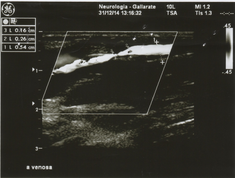
Here, we report a case of a 38-year-old patient, who underwent hemodialysis firstly by a CVC long-term into right jugular vein and then by a prosthetic fistula in the ipsilateral limb. The patient presented many episodes of vascular access thrombosis that required endovascular interventions. The ultrasound screening and CT-angiography revealed an asymptomatic stenosis of the superior cava vein, which treatment with the implantation of vascular stent resulted in an initial improvement of vascular access performance. However, in the following months, a restenosis was observed that required new interventions to reestablish a satisfactory vascular access function.
This case highlights that patients on hemodialysis should undergo proper clinical and instrumental follow-up in order to prevent or early recognize vascular access complications.
KEYWORDS: echocolordoppler, hemodialysis, vascular access, graft.