The renal lesions in Bardet-Biedl Syndrome: history before and after the discovery of BBS genes

Abstract

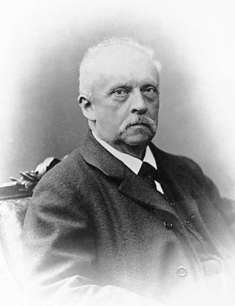
Various renal lesions of the Bardet-Biedl syndrome (BBS) have been described, including macroscopic and microscopic kidney abnormalities, polyuria, polydipsia and chronic renal failure. However, these renal symptoms were completely overlooked for about fifty years after the first description of the syndrome. The observation of a familial origin of the syndrome began in 1753, with Maupertuis and Réaumur describing hereditary forms of polydactyly. In the early 19th century, Martin mentioned an inherited case of blindness. Subsequently, von Graefe (1858) reported on a familial occurrence of both of blindness and deafness. The introduction of the ophthalmoscope by von Helmholtz (1851) allowed for the identification of patients with retinal degeneration. Systematically using this instrument, Laurence and Moon (1866) were the first to describe a familial case of retinal degeneration combined with obesity and cognitive impairment. Due to the influential work of Froehlich, Cushing, and Babinski, attention then shifted to obesity. The syndrome was definitively identified by 1920 through Bardet’s observations familial cases of obesity, blindness, polydactyly, and hypogonadism. Biedl in 1922 observed further cases of the syndrome. In recognition of this history, the disease was named Laurence-Moon-Bardet-Biedl Syndrome. The renal anomalies were not described until fifty years later, in 1977. In 1993, the quest for the genes involved in BBS began with the isolation of 21 different genes. In 2003 two concepts emerged: the existence of a spectrum of ‘ciliopathies’ and the concept of the BBSome. Afterwards, the gene-phenotype relationship was researched using transgenic mice.

Keywords: ciliopathies, hereditary, obesity, retinitis, chronic kidney disease

Ci spiace, ma questo articolo è disponibile soltanto in inglese.

INTRODUCTION

According to the influential theory of Thomas Kuhn (1922-1996) (1), most scientists work constrained by current influential paradigm and are devoted to solving small problems (‘puzzle-solving’). The dominant paradigm is important for the interpretation of the data, but it may blind scientists to new phenomena not considered part of the paradigm. One example of this theory comes from the field of nephrology, where the pivotal renal anomalies in Bardet-Biedl Syndrome went completely unnoticed for more than 50 years after the discovery of the syndrome. Tus, the BBS syndrome is an example of how an essential clinical element may go unnoticed for a long time and is evaluated only after a shift in the attention of the scientific community (specifically, the introduction of renal biopsy and immunofluorescence).

The Bardet-Biedl Syndrome (BBS) is a rare genetic disorder characterized by retinal degeneration, polydactyly, obesity, learning disabilities, hypogonadism and renal anomalies. Various renal lesions of BBS have been described including (i) fetal lobulation (ii) calyceal clubbing, (iii) focal sclerosing glomerulonephritis, (iv) interstitial nephritis, and (v) changes in the glomerular basement membrane. Polyuria, polydipsia and chronic renal failure have been also reported in many case reports (2). Although the renal anomalies are today one of the primary features of the disease, it took almost 50 years after the description of the syndrome for renal symptomatology to be included.

Here we will review the observations that drew the attention of Bardet and Biedl to the disease and why the renal features were not observed. Afterwards, we will focus on the role that the identification of BBS genes played in changing our perception of the disease and its renal lesions. A timetable of the discoveries is summarized in Table 1.


HOW THE SYNDROME WAS DISCOVERED

The identification of BBS required the evolution of the following concepts: 1) the existence of hereditary forms of blindness and polydactyly, which fostered the search for combined hereditary forms of more complex diseases 2) the invention of the ophthalmoscope, which allowed scientists to identify and classify retinal degeneration and 3) a paradigm-shift concerning the nature of obesity, which focused attention on hereditary forms of obesity (such as BBS), but also served as a blinder impeding the identification of other features such as kidney failure.

The observation of a familial origin of the syndrome began in 1753, with Maupertuis and Réaumur (Figure 1, Figure 2) describing hereditary polydactyly. While polydactyly was widely known since ancient times, the hereditary aspect of the malformation gained notice in the late 1700s. Pierre-Louis Moreau de Maupertuis, (born Sept. 28, 1698, Saint-Malo, France—died July 27, 1759, Basel, Switz.), was a mathematician and astronomer who popularized Newton’s theories (3). In Système de la nature ou Essai sur les corps organisés (1751) he studied the transmission of polydactyly in four generations of a Berlin family, providing the first report of the trait as hereditary (4). Renè-Antoine Ferchault de Réaumur (1683-1757), the famous French scientist who gave his name to the temperature scale, is reported by Huxley (1894-1963) (5) to have analyzed data from three families (named Kelleia) from Malta with hereditary polydactyly. Similar to polydactyly, progressive blindness was also known since ancient times; however, the possibility of a hereditary form of blindness was first noted in the early 19th century by Martin. He reported, in the Baltimore Medical and Physical Recorder (1809), on the Lecomptes, a Maryland family of French origin whose members suffered progressive blindness (5). While none of these authors were describing actual cases of BBS, their work did refocus subsequent researchers on hereditary forms of polydactyly and blindness.

Indeed, soon after, Albrecht von Graefe (1828-1870) (6) and thereafter Liebreich first reported a hereditary combination of blindness and deafness in cases of what would be called retinitis pigmentosa, furthering the concept of combined forms of hereditary traits, and these observations are, in fact, cited by Laurence and Moon in their work (see below). Another essential discovery that must be acknowledged for the history of BBS was the invention of the ophthalmoscope in 1851 by Hermann von Helmholtz (1821-1894), which allowed the observation of the retina and hence the definition of retinitis pigmentosa (Figure 3). The use of the new device, the ophthalmoscope, was hence promoted in England by John Zachariah Laurence (1829-1870), a surgeon and ophthalmologist at the ophthalmologic hospital in Southwark (Figure 4). In 1866, together with his colleague Robert Charles Moon (1844-1914) (Figure 5), a house surgeon at the same hospital (who then moved in Philadelphia), they were the first to describe, using the ophthalmoscope, a familial case of combined retinal degeneration, obesity, and cognitive impairment (7).

In the first years of the 20th century, medical attention shifted to hypothalamic forms of obesity-hypogonadism thanks to the work of a neurologist, Joseph Babinski (1857-1932), a pharmacologist, Alfred Fröhlich (1871-1953) (8) and a neurosurgeon, Harvey Cushing (1869-1939) (9). Again, in the history of science, we see how important advances in one field may come through collaborations with other fields, and how this chance partnership was a necessary step in fully defining BBS. Fröhlich’s strong influence is visible when the first report of a BBS case was attributed to a pituitary malfunction.

Around this period a certain number of observations of obesity, polydactyly and retinitis pigmentosa are reported by several authors: in 1887 Ferdinand-Jean Darier (1856-1938) reports the association of retinitis pigmentosa and polydactyly (10). In 1989 Elie von Cyon (also known as de Cyon, 1843-1912) presents the case of a 12-year old boy with obesity, growth and mental retardation, and hereditary polydactyly (11). In 1898 Ed Fournier reports retinitis pigmentosa and syndactyly (12). In 1913 Rozabel Farnes reports adipose-genital syndrome with polydactyly (13). In 1914 an Italian radiologist working in Naples, Mario Bertolotti (1876-1957) presented the case of Marguerite Catt, 39 years old, with polydactyly, mental retardation, obesity, retinitis pigmentosa, and hypogonadism (14). In 1918 J Madigan and Thomas Verner Moore (1877-1969) described a case of mental retardation, obesity, hypogonadism, retinitis pigmentosa, and tapering toes (15).
Finally, in 1920 a French medical student, George Louise Bardet (1885-1966), in his medical degree thesis, collected all these cases and his own observation of a familial case of obesity, hexadactyly, retinitis pigmentosa and hypogonadism and proposed the existence of a triad (13). He discussed this finding using the current paradigm of hypophyseal/hypothalamic obesity: “Two congenital malformations (hexadactyly and retinitis pigmentosa) in a child who became obese from birth. What is the gland which can be incriminated? (…) We believe this case must be attached to a very special clinical variety of hypophysis obesity”. Bardet’s triad (obesity, polydactyly, retinitis pigmentosa) gained success after the father of modern endocrinology, Arthur Biedl (1869-1933), in 1922 observed further cases of the syndrome. Biedl named the syndrome adipose-genital dystrophy and thought it was of cerebral origin, in line with the paradigms of that period (Figure 6). In recognition of this history, the disease was named Laurence-Moon-Bardet-Biedl Syndrome. Later, thanks to the work of Ammann in 1970 and Schachat and Maumenee in 1982, Laurence-Moon and Bardet-Biedl Syndromes came to be considered two different entities and possibly part of the same disease spectrum. In the first half of 1900, BBS was officially defined, but none of these authors noticed abnormalities in kidney function, which is today acknowledged as an important signature of the syndrome.
Why then were the renal features of the syndrome missed for almost 50 years? It is tempting to see this as an example of Kuhn’s hypothesis that scientists work on ‘puzzle-solving’ within an influential paradigm. The paradigm of that period was hypothalamic obesity, whereas kidney failure was not considered. Scientists observing new cases of BBS focused on obesity and dismissed other possible features of the disease.
It is intriguing that, even in 1995, in the excellent editorial by George Bray (born 1931) on the syndrome in Obesity Research, kidney dysfunction is completely ignored by the author (16).


THE RENAL LESIONS BEFORE BBS GENES

Awareness of the renal involvement in BBS starts in the late 1960s with the work of McLoughlin and Shanklin (17), Nadjmi (18), Hurley (19) and Falkner (20). McLoughlin and Shanklin (17), Nadjmi et al. (18) first reviewed necropsies of BBS from the literature and found a high incidence of renal/genitourinary malformations; Nadjmi further observed that most of cases reported in the literature since 1940 died for uremia and therefore renal failure was a major cause of early death in BBS patients. According to Nadjmi, the first autopsy reporting a BBS subject passed due to uremia was by Radner in 1940 (Acta Med Scand 105:141); however, genitourinary tract malformations were already observed since 1938 by Griffiths (J Neurol Psychiat 1:1-6), and Riggs (Arch Neurol Psychiat 39:1041). It is possible that the systematic renal involvement in BBS was missed before because the histologic classification of kidney diseases reached its maturity only when kidney biopsy and the kidney immunofluorescence have been available around 1950, thus driving attention to this organ.

The diffusion of the technique of percutaneous kidney biopsy by Nils Alwall (1904-1986) allowed Hurley et al (19) to first report histological data from a series of nine BBS children (Figure 7 A-B). The results were quite variable, from mesangial proliferation to sclerosis, cystic dilatation of the tubules, cortical and medullary cysts, periglomerular and interstitial fibrosis, chronic inflammation.

Falkner et al. (20) found in a 24-month old child with BBS right sided vesical-ureteral reflux, cystocele, urinary tract infections, growth arrest of the right kidney. They also confirm the mesangial hypercellularity by percutaneous biopsy (Figure 7 C).

In 1990 the incidence of renal abnormalities in BBS was finally determined to be very high: up to 90% of the patients, and therefore become a new signature of the syndrome, more than 50 years from its initial definition (2). In the meanwhile, the spectrum of renal abnormalities was stably defined as:

Functional: polyuria, polydipsia, aminoaciduria, reduction of maximum concentrating capacity, chronic renal failure, hypertension

Macroscopic: fetal lobulation, cystic dysplasia and calyceal cysts, small kidneys, calyceal clubbing or blunting

Microscopic: swelling of endothelial cells, tubular and interstitial nephritis with glomerulosclerosis.

In conclusion, we believe that the attention to the nephrological character of the BBS was finally reached only when (i) technical advancements were available (that is the invention of the percutaneous biopsy) and (ii) when a general attention of the medical entourage was driven towards the kidney function: we should remind that in 1943 Willem Johan Kolff (1911 – 2009) first built a dialyzer machine, further developed by Nils Alwall. At the end of 60’ nephrology was a mature science and the greater awareness towards uremia led to a revision of syndromic diseases.

However, the condition remained largely unclear even after the discovery of the renal abnormalities: major advances in a new behind the complex trait was the discovery of the gene defects causing BBS.


THE RENAL LESIONS AFTER BBS GENES

The quest for the genes occurred in two phases: from 1993 to 2000 a genetic mapping was pursued, with the identification of several DNA loci involved in the disease. In 2000 the identification of the first BBS gene (now they number 21), MKKS, based on the similarity between the BBS and the McKusick-Kaufman syndrome (MKS), occurred (21). In 2003 Ansley et al demonstrated that mammalian BBS8 gene was restricted to ciliated cells (21). This finding raised the hypothesis that BBS proteins play a role in cilia function. Meanwhile, other genes of the same family were found to cause BBS, with at least 17 different genes implicated up to now.

The field was quite mature at the time because a second, more common condition, was already found to involve cilia: the polycystic kidney disease (PKD). This is also a hereditary condition and followed almost the same path of BBS (anatomical period-genetic period-functional period), which ultimately led to the paradigm of the involvement of cilia dysfunction in the genesis of the disease.

It should be stressed that, again, the major advancement in the paradigm did not come directly from the studies on the disease, but from studies on flagellated protozoa: it was a genetic study on immobile forms of these protozoa which led to the identification of this gene. When the same was found to be involved in PKD and then in other diseases such as BBS, it was almost immediate the formation of a new paradigm of ‘ciliopathies’. All genes involved in these genetic diseases and in the cilium were then functionally grouped in a multiprotein complex called BBSome.

After the period of discovery of BBS genes and the construction of the concept of the BBSome, some new insights in the renal pathology of BBS have been addressed. First, the gene-phenotype relationship has been studied in much detail, with a categorization of mutations leading to various associations of the visual, metabolic and kidney phenotypes (23, 24). Second, a number of transgenic mice are now available for testing of pathogenic hypotheses and new pharmacological approaches. Risk factors for the development of the renal disease have been studied in large cohorts (22 – 24), and the usefulness of renal transplantation has been demonstrated in a separate study (25, 26). A contribution for low protein diet in the preservation of renal function in BBS has also been reported (27). Finally, a study from one of us (28, 29) showed combined impaired water handling in BBS.

These functional changes in BBS kidney might be mediated, at least in part, by mistrafficking of apical membrane proteins, leading to tubular dysfunction (41). In turn, this might be related to the renal hyposthenuria in BBS, that has been recognized as the most common renal dysfunction in the absence of renal insufficiency (42, 43).


Acknowledgments

I am indebted with dr. David Widmer, who critically reviewed the manuscript, with useful suggestions and critiques.


REFERENCES
  1. Kuhn TS. The Structure of Scientific Revolutions. Chicago, IL: University of Chicago Press; 1996.
  2. Regenbogen L, Eliahou H. Diseases affecting the eye and the kidney. Karger; 1993.
  3. The Editors of Encyclopædia E. Encyclopaedia Britannica. Pierre-Louis Moreau de Maupertuis (Internet). 1998.. Available from: https://www.britannica.com/biography/Pierre-Louis-Moreau-de-Maupertuis
  4. Cobb M. Hereditary before genetics: a history. Nat. Rev Genet. 2006;7:953–8.
  5. Harper P. A short history of medical genetics. Oxford press; 2008.
  6. von Graefe A. Vereinzelte Beobachtungen und Bemerkungen: Exeptionelles Verhalten des Gesichtfeldes bei Pigmentartung der Netzhaut. Arch. für Klin. und Exp. Ophthalmol. 1858;4:250–3.
  7. Laurence J, Moon RC. Four cases of retinitis pigmentosa occurring in the same family accompanied by general imperfection of development. Ophthalmic Rev. 1866;2:32–41.
  8. Fröhlich A. Ein Fall von Tumor der Hypophysis cerebri ohne Akromegalie (A case involving a tumor of the hypophysis cerebri without acromegaly). Wiener Klin. Rundschau. 1901;15:833–6; 906–8.
  9. Cushing H. The basophil adenomas of the pituitary body and their clinical manifestations (pituitary basophilism). Bull. Johns Hopkins Hosp. 1932;50:137–95 Reprinted in: Ann R Coll Surg Engl. 44.
  10. Darier F-J. Observation VI. Ophtalmol. Arch. 1887;174. Cited in: Bardet G, 1920.
  11. De Cyon E.Traitement de l’acromégalie par l’hypophysine. Bull. De l’Acad. de Med. Paris. 1898; 11:444. Cited in: Bardet, 1920.
  12. Fouriner E. case #328. These de Paris; 1898. Cited in: Bardet, 1920.
  13. Bardet G. Sur un Syndrome d ’Obésité Congénitale avec Polydactylie et Retinite Pigmentaire (Contribution a l’etude des formes clinique de l’Obésité hypophysaire). Thése Paris No 470. Paris: Translated in: Obesity Res 1995 3:387; 1920.
  14. Bertolotti M. Polydactyly arrested in development of members and concomitant hypophysis dystrophy. J. Acad. Med. Torrino. 1914;6.
  15. Madigan JJ., Moore TV. DYSTROPHIA ADIPOSOGENITALIS (FRÖHLICH’S SYNDROME)REPORT OF CASE. J. Am. Med. Assoc. 1918;70:669-71. Cited in Bardet, 1920.
  16. 16. Bray G. Laurence Moon Bardet and Biedl: reflections on a syndrome. Obes Res. 1995;3:383–6.
  17. McLoughlin T, Shanklin D. Pathology of Laurence-Moon-Bardet-Biedl syndrome. J. Pathol. Bacteriol. 1967;93:65–79.
  18. Nadjmi B, Flanagan M, Christian J. Laurence-Moon-Biedl syndrome associated with multiple genito-urinary tract anomalies. Am J Dis Child. 1969;117:352–6.
  19. Hurley R, Dery P, Nogrady M, Drummond K. The renal lesion of the Laurence-Moon-Biedl syndrome. J Pediatr. 1975;87:206–9.
  20. Falkner B, Langam C, Katz S. Renal histopathological changes in a child with Laurence-Moon-Biedl syndrome. J Clin Pathol. 1977;30:1077–81.
  21. Lupski JR, Katsanis N, Beales PL, Woods MO, Lewis RA, Green JS, et al. Mutations in MKKS cause obesity, retinal dystrophy and renal malformations associated with Bardet-Biedl syndrome. Nat. Genet. 2000;26:67–70.
  22. König J, Kranz B, König S, Schlingmann KP, Titieni A, Tönshoff B, et al. Phenotypic Spectrum of Children with Nephronophthisis and Related Ciliopathies. Clin. J. Am. Soc. Nephrol. 2017;CJN.01280217.
  23. Esposito G, Testa F, Zacchia M, Crispo AA, Di Iorio V, Capolongo G et al.. Genetic characterization of Italian patients with Bardet-Biedl syndrome and correlation to ocular, renal and audio-vestibular phenotype: identification of eleven novel pathogenic sequence variants. BMC Med. Genet. 2017;18(1):10.
  24. Stoetzel C, Laurier V, Faivre L, Mégarbané A, Perrin-Schmitt F, Verloes A, et al. BBS8 is rarely mutated in a cohort of 128 Bardet–Biedl syndrome families. J. Hum. Genet. 2006;51:81–4.
  25. Hooda A, Karan S, Bishnoi J, Nandwani A, Sinha T. Renal transplant in a child with Bardet-Biedl syndrome: A rare cause of end-stage renal disease. Indian J. Nephrol. 2009;19:112.
  26. Sharifian M, Dadkhah-Chimeh M, Einollahi B, Nafar M, Simforoush N, Basiri A, et al. Renal transplantation in patients with Bardet-Biedl syndrome. Arch. Iran. Med. 2007;10:339–42.
  27. Dervisoglu E, Isgoren S, Kasgari D, Demir H, Yilmaz A. Obesity control and low protein diet preserve or even improve renal functions in Bardet-Biedl syndrome: a report of two cases. Med. Sci. Monit. 2011;17:CS12–4.
  28. Zacchia M, Zacchia E, Zona E, Capolongo G, Raiola I, Rinaldi L, et al. Renal phenotype in Bardet-Biedl syndrome: a combined defect of urinary concentration and dilution is associated with defective urinary AQP2 and UMOD excretion. Am. J. Physiol. – Ren. Physiol. 2016;311:F686–94.
  29. Zacchia M, Di Iorio V, Trepiccione F, Caterino M, Capasso G. The Kidney in Bardet-Biedl Syndrome: Possible Pathogenesis of Urine Concentrating Defect. Kidney Dis. 2017;3:57–65.
  30. Gourdol O, David L, Colon S, Bouvier R, Ayral A, Aguercif M, et al. Renal involvement in the Laurence-Moon-Bardet-Biedl syndrome. Apropos of 3 cases. Pediatrie. 1984;39:175–81.
  31. Kwitek-Black AE, Carmi R, Duyk GM, Buetow KH, Elbedour K, Parvari R, et al. Linkage of Bardet-Biedl syndrome to chromosome 16q and evidence for non-allelic genetic heterogeneity. Nat. Genet. 1993;5:392–6.
  32. Leppert M, Baird L, Anderson KL, Otterud B, Lupski JR, Lewis RA. Bardet-Biedl syndrome is linked to DNA markers on chromosome 11q and is genetically heterogeneous. Nat. Genet. 1994;7:108–12.
  33. Sheffield VC, Carmi R, Kwitek-Black A, Rokhlina T, Nishimura D, Duyk GM, et al. Identification of a Bardet-Biedl syndrome locus on chromosome 3 and evaluation of an efficient approach to homozygosity mapping. Hum. Mol. Genet. 1994;3:1331–5.
  34. Carmi R, Rokhlina T, Kwitek-Black AE, Elbedour K, Nishimura D, Stone EM, et al. Use of a DNA pooling strategy to identify a human obesity syndrome locus on chromosome 15. Hum. Mol. Genet. 1995;4:9–13.
  35. Pazour GJ, Dickert BL, Vucica Y, Seeley ES, Rosenbaum JL, Witman GB, et al. Chlamydomonas IFT88 and its mouse homologue, polycystic kidney disease gene tg737, are required for assembly of cilia and flagella. J. Cell Biol. 2000;151:709–18.
  36. Ansley SJ, Badano JL, Blacque OE, Hill J, Hoskins BE, Leitch CC, et al. Basal body dysfunction is a likely cause of pleiotropic Bardet-Biedl syndrome. Nature. 2003;425:628–33.
  37. Blacque OE, Reardon MJ, Li C, McCarthy J, Mahjoub MR, Ansley SJ, et al. Loss of C. elegans BBS-7 and BBS-8 protein function results in cilia defects and compromised intraflagellar transport. Genes Dev. 2004;18:1630–42.
  38. Moore SJ, Green JS, Fan Y, Bhogal AK, Dicks E, Fernandez BA, et al. Clinical and genetic epidemiology of Bardet-Biedl syndrome in Newfoundland: a 22-year prospective, population-based, cohort study. Am. J. Med. Genet. A. 2005;132A:352–60.
  39. Badano JL, Mitsuma N, Beales PL, Katsanis N. The Ciliopathies: An Emerging Class of Human Genetic Disorders. Annu. Rev. Genomics Hum. Genet. 2006;7:125–48.
  40. Seo S, Baye LM, Schulz NP, Beck JS, Zhang Q, Slusarski DC, et al. BBS6, BBS10, and BBS12 form a complex with CCT/TRiC family chaperonins and mediate BBSome assembly. Proc. Natl. Acad. Sci. U. S. A. 2010;107:1488–93.
  41. Zacchia M, Esposito G, Carmosino M, Barbieri C, Zacchia E, Crispo AA et al. Knockdown of the BBS10 Gene Product Affects Apical Targeting of AQP2in Renal Cells: A Possible Explanation for the Polyuria Associated with Bardet-Biedl Syndrome. Genet Syndr. Gene Ther 2014; 5:3.
  42. Zona E, Zacchia M, Di Iorio V, Capolongo G, Rinaldi L, Capasso G. Patho-physiology of renal dysfunction in Bardet-Biedl Syndrome. G Ital Nefrol 2017; 34: 62-72.
  43. Zacchia M, Capolongo G, Trepiccione F, Marion V. Impact of Local and Systemic Factors on Kidney Dysfunction in Bardet-Biedl Syndrome. Kidney Blood Press Res. 2017 ;42:784-793.
  44. Sorsby A. John Zachariah Laurence-A belated tribute. Br. J. Ophtalmol. 1932; 16:727-40.